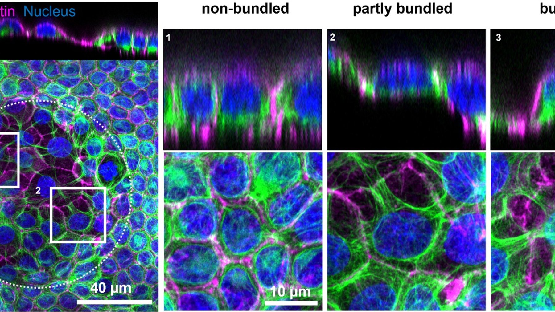
How do living tissues endure a pull that lasts not just seconds, but hours or even days? New research led by the Institute for Bioengineering of Catalonia (IBEC) shows that epithelial cells do not simply brace against the strain. They gradually rebuild their internal keratin scaffolding across neighboring cells, while their nuclei become detached from the protective mesh that normally surrounds them.
The discovery reveals a previously unknown way tissues adapt to sustained mechanical forces. It could help researchers better understand processes ranging from embryonic development to the repeated expansion and contraction of organs such as the bladder and mammary gland.
Keratin does not react all at once
Keratin is a major part of the cytoskeleton, the network of protein filaments that gives cells their shape and mechanical strength. Scientists already knew that keratin helps tissues withstand large deformations, but its response to long-lasting stretching had remained unclear.
To investigate, the team combined engineered epithelial tissues with a custom-built microfluidic stretching device, live-cell microscopy, and multiscale computer modeling. Rather than seeing an immediate response, the researchers watched individual keratin filaments reorganize slowly over several hours into thick, star-shaped bundles.
Those bundles did not stop at the border of a single cell. They extended between neighboring cells and formed shared, supracellular networks across the tissue, a bit like separate support beams being joined into one larger frame.
The rebuilding process pushes nuclei out of their keratin cages
The structural shift began at junctions where three cells meet. Keratin filaments gradually disappeared from those regions and accumulated into thicker bundles, first in a few isolated cells and then in expanding clusters connected by a common network.
Computer simulations suggested that the same forces responsible for creating the bundles also pushed each nucleus away from the keratin cage around it. “The simulation of intracellular dynamics revealed that the same process that forms these striking bundles also generates forces that ultimately push the nucleus out of its cage,” said Marco Pensalfini of Queen Mary University of London.
Live imaging backed up that prediction. As the bundles thickened, the nucleus slowly detached from the surrounding keratin mesh until only a small amount of filament remained connected to it. The researchers described this unexpected event as “nuclear uncaging.”
Does nuclear uncaging protect cells or expose them?
That question remains open. Removing the keratin cage could leave the nucleus more directly exposed to mechanical forces, which may make it more vulnerable during prolonged stretching.
On the other hand, disconnecting the nucleus from a highly stressed cytoskeletal network could reduce the amount of force transmitted to it. “One of the most interesting questions raised by our study is whether this release process ultimately protects the nucleus or makes it more vulnerable,” said Xavier Trepat, an ICREA research professor at IBEC and co-senior author of the study.
Future work will need to determine which effect dominates. For now, the study shows that what looks like a simple stretch from the outside can trigger a much more complex mechanical reorganization inside the tissue.
Actin helps control the pace of change
The researchers also found that actin, another major component of the cytoskeleton, plays an important regulatory role. When they weakened the molecular links between actin and keratin, the formation of keratin bundles sped up by almost three times.
In practical terms, the two filament systems appear to work together to control how quickly tissue architecture changes under sustained strain. Keratin provides much of the structural reinforcement, while its connections with actin help govern when and how that reinforcement is rebuilt.
That coordination matters because cells within a tissue must adapt together. Think of a building where the beams and joints cannot respond independently when the whole structure is under a repeated load.
Why prolonged stretching matters in the body
Similar mechanical conditions arise during embryonic development and in organs that repeatedly expand and contract. The bladder fills and empties, while mammary tissue changes substantially over time, placing cells under forces that can last far longer than a quick tug in a laboratory.
“These findings may help us better understand how tissues adapt to prolonged stretching in a wide range of biological contexts,” said Tom Golde, a postdoctoral researcher in Trepat’s group and first author of the study. He added that the mechanism may also be relevant to diseases in which the keratin network is disrupted.
The engineering comparison is hard to miss. Bridges and buildings are reinforced so they can survive repeated loads, and cells appear to use a related strategy by continually rebuilding their internal scaffolding. The twist is that strengthening the tissue may also change how the nucleus is mechanically connected.
A new view of tissue resilience
The study does not yet establish whether nuclear uncaging is beneficial or harmful. What it does show is that tissue resilience is an active, collective process rather than a passive property of tough cellular material.
As stretching continues, cells remodel keratin across their neighbors, adjust their relationship with actin, and alter the physical protection around their nuclei. That is a major shift in how scientists can think about the mechanics of tissues under everyday biological strain.
The study was published in Nature Physics.