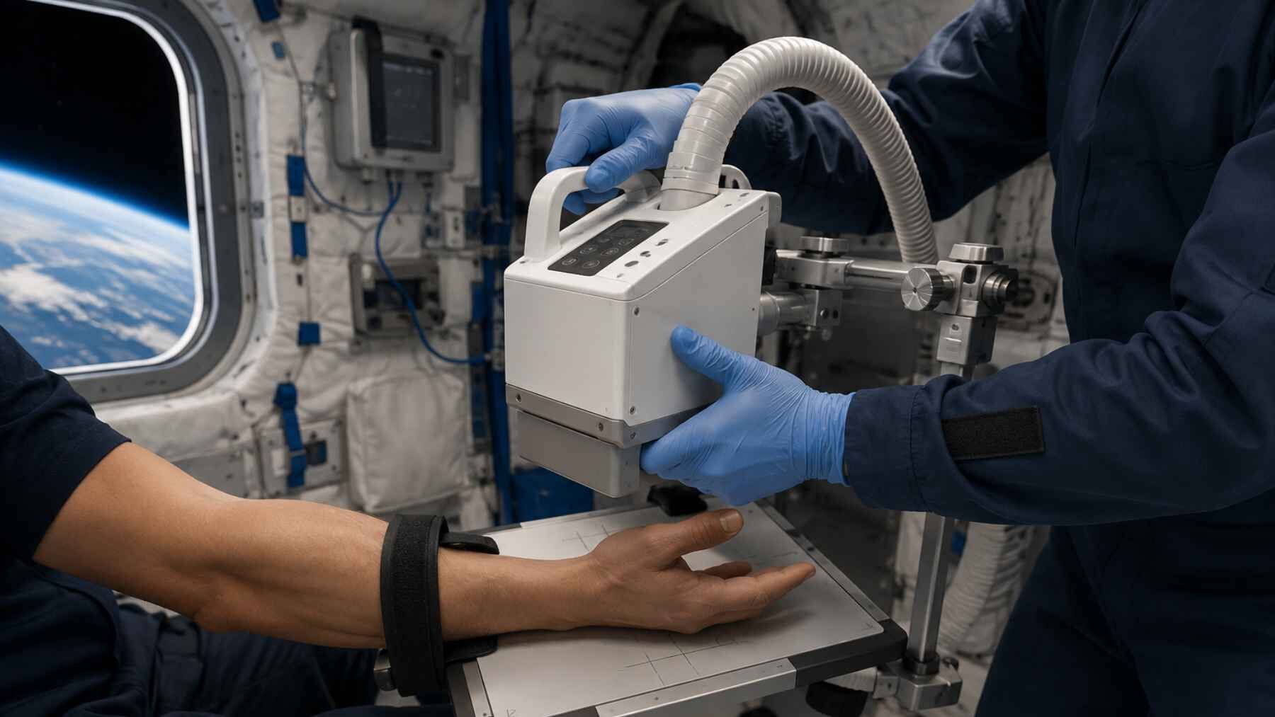
What happens if an astronaut suffers a serious fall far from the nearest hospital? A small portable X-ray machine has now passed a test that aerospace doctors have waited decades to see, producing the first diagnostic human radiographs during an orbital flight. The result could give future crews heading toward the Moon a much clearer way to assess suspected fractures and other medical problems.
The experiment also carries an Earthbound promise. A device light and simple enough for nonmedical crew members to use in microgravity could eventually support rescue teams, rural clinics, and communities located many hours from a major imaging center. The breakthrough is not a finished lunar emergency room, but it shows that the basic idea works.
A medical first aboard Fram2
The test took place during Fram2, a private SpaceX mission that launched on March 31, 2025, and spent 3 days and 14 hours in a polar orbit roughly 264 to 280 miles above Earth. Three crew members received four hours of training, while orbital time limits meant that only two underwent in-flight human imaging. With no live ground assistance, the crew obtained X-rays of a hand, forearm, chest, abdomen, and pelvis, along with a calibration object and a smartwatch.
For more than four decades, ultrasound has been the only reliable medical imaging method used during spaceflight. It is useful and does not expose a patient to ionizing radiation, but it depends heavily on probe placement, operator skill, and sound transmission through the body. X-rays can answer different questions, especially when doctors need to look at bones or examine structures hidden inside equipment.
Sheyna Gifford, an aerospace medicine physician at Mayo Clinic and the study’s lead researcher, summed up the appeal in simple terms. “X-rays are fast, easy and diagnostically valuable,” she said. That sounds obvious in a hospital, but making the same technology work while the patient, detector, and operator are all floating is another matter.
The images passed the test
Three independent radiologists evaluated the preflight and in-flight images for overall quality, spatial detail, contrast, and positioning. The seven radiographs taken in orbit received an average overall quality score of 4.86 out of 5, compared with 5 before launch. Every image still reached the study’s threshold for diagnostic quality.
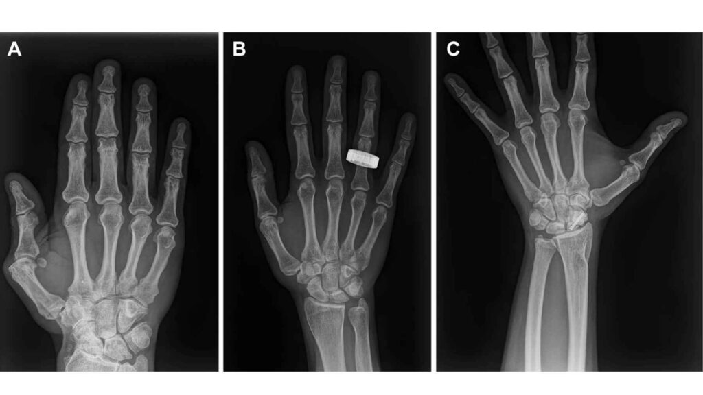
The biggest weakness was positioning, not sharpness. For chest, abdomen, and pelvis images, the average positioning score fell from 4.95 before flight to 4.07 in orbit, a reminder that even a straightforward hospital procedure becomes awkward when nothing stays put. Crew members asked for better clamps, mounting points, or hook-and-loop fasteners to secure the generator and detector.
The machine also survived the trip home. Its exterior suffered bent guards and a broken plastic part during landing and recovery, yet its internal hardware and X-ray output continued to function normally. Estimated exposure from the system was no greater than that associated with standard clinical imaging on Earth.
More than a medical scanner
The experiment was designed to look inside machines as well as people. In orbit, the system revealed internal components in test objects at scales smaller than roughly 0.04 inch, without taking them apart. That type of nondestructive inspection could help crews examine electronics, satellite parts, or the layered components inside a spacesuit.
That matters because a damaged suit or electrical unit cannot always be dismantled safely during a mission, and replacement parts may be days or months away. A compact radiography system could act like a mechanic’s flashlight, showing what is happening beneath the surface before a crew makes a risky repair.
Researchers also see potential for X-ray devices on lunar rovers that could help analyze the Moon’s surface. One compact system might support astronaut health, spacecraft maintenance, and scientific work. That is valuable when every pound of cargo and every watt of power must earn its place.
The Earthbound payoff
Portable X-ray units are already used at sporting events and in low-resource settings, and some can run on solar power. Their digital images appear immediately on a computer, removing the need to develop film and making remote review possible. For a rural clinic or rescue team working in a tight space, that could mean getting useful information before arranging a long and expensive transfer.
The limits of a small first study
Still, this was a small feasibility study, not a hospital trial. The participants were healthy, the number of images was limited, no actual injury was diagnosed, and the radiographs were reviewed on Earth rather than used for real-time treatment decisions. Longer missions will also require clear radiation rules, better positioning procedures, and dependable remote interpretation.
The next version will need to be smaller, tougher, and easier to secure inside a spacecraft, perhaps with automated guidance for poorly aligned images. The first orbital X-rays answered whether this can be done, and now comes the harder work of making it routine.
The full study was published in Radiology.
Most read
- US scientists poured 16,500 gallons of lye into the Gulf of Maine and dyed the water bright pink to track it, the first federally approved try at reversing ocean acidity
- Soviet scientists released giant red king crabs into the Barents Sea in the 1960s to build a fishery, but decades later those invaders are still changing the seafloor
- New Orleans is collecting thousands of used Christmas trees and dropping them from helicopters to slow the loss of land, which has already exceeded 5,000 square kilometers
- The U.S. will pay up to $65 million to leave Colorado River water in Lake Mead, and Hoover Dam explains why
- Britain is dropping 20 concrete reef cubes and 44 tons of scallop shells into the sea, and the goal is to bring back oysters down more than 95% since the 1800s